Q&A with Paola Marino
Microscopes allow researchers to visualize seemingly invisible things. But practical purposes aside, these glimpses into the microscopic world often look like works of art. For Paola Marino, conducting lab work is very much an artistic experience. A Master of Science candidate at Concordia University, Paola is studying the design, synthesis, and functionalization of metal–organic frameworks for ophthalmic drug delivery.
Paola was a 2020 Visualizing Science Contest participant whose image was selected for the 2021 cover of the Canadian Journal of Chemistry.
Where do you find inspiration for your microscopy work?
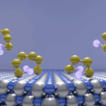
When it comes to inspiration for my microscopy work, there are several reoccurring concepts that come to mind. These concepts all have an underlying connection to the universal idea of art, and more specifically, I gain inspiration from visual elements such as patterns, shapes, colors, lines, and textures. Whenever I take a trip to the museum, I enjoy analyzing artwork, not only for the visual elements, but also, for the emotions that come alive while analyzing a certain piece. A year ago, I attended an incredible and mesmerizing exhibit, “Imagine Van Gogh” that had huge projections of his paintings, and one that quickly captivated my attention was his painting entitled “The Starry Night”. For my image “Metal–Organic Galaxy”, that is currently featured on the 2021 cover of the Canadian Journal of Chemistry (CJC), it was a direct tribute to the painting that fascinated me during my visit to the art center.
When your sample matches the colour of your gloves🧤#chemistry #MOFs #research pic.twitter.com/ooKGkCGCiW
— Paola Marino (@paolamchem) June 17, 2021
What kind of tools do you use for image processing?
For image processing, I use Adobe Photoshop which serves as a colorizing tool for the images I collect. Since the scanning electron microscope (SEM) images are captured in black and white, I like to add color to my image to create a story that showcases my inspiration. The tools that Photoshop provides allow me to enhance certain areas, elements, and shapes of my images with various textures, dimensions, and contrasts. In relation to the artwork by Van Gogh entitled “The Starry Night”, the sky was a deep blue and the stars punctured through the sky in bright yellow tones. The clusters of metal–organic frameworks (MOFs) seen on the CJC cover, which I otherwise coined as octahedral stars, are reminiscent of this painting that I colorized with various shades of blue and yellow. I appreciate exploring and learning about art, and then determining how I can apply it to my work in the laboratory.
Tell us about a time when taking an image with the microscope didn’t go as planned
Working with a SEM can result in some unexpected moments. One time in particular would be when I placed a sample for analysis and after adjusting the settings, the accelerating voltage exposed to my uncoated microcrystalline MOF sample suddenly triggered the particles to burst. This resulted in some particles disconnecting from their initial form and transforming into something unique. That was the first time I witnessed such a phenomenon under the microscope, and it reminded me how chemistry can be unpredictable.