Q&A with Dr. Christian Lacroix
It’s been the “Year of the Hobby”. Gardening being one of many pastimes taken up during our time at home. Perhaps you spent a small fortune on succulents at your local greenhouse or find yourself caked in topsoil daily as you nurture your herbs. You now consider yourself a bonafide plant expert. But there’s always more to learn, isn’t there?
In this Q&A with Dr. Christian Lacroix, Co-Editor of Botany and previous judge of the Visualizing Science Contest, we get a glimpse into the world of plant development. Through the use of scanning electron microscopy, he captures plant development in astounding detail, showcasing the beautiful structural complexity that lays within flowers.
What drew you to plant science?
When I was an undergraduate student at McGill University, I took an elective course in plant anatomy from Dr. Rolf Sattler. His enthusiasm for the subject matter and his approach to teaching connected with me and I haven’t looked back since. In subsequent years, I was able to do a directed studies under his supervision and eventually completed my M.Sc. in his laboratory.
What is your all-time favourite visualization of plant development?
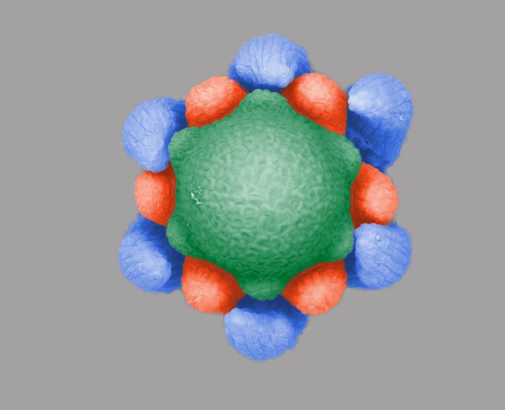
As I developed my knowledge base on plant development, I became very familiar with the technique of scanning electron microscopy (SEM). The use of SEM has revolutionized our ability to examine the detail of various types of surfaces, and in my case how organs actually form on the shoot apical meristem of plants. My all-time favourite images are those that feature this unique structure in plants, responsible for producing a variety of organs like leaves and flowers. The photo [below] features a top view of the shoot apical meristem of Myriophyllum aquaticum; the central dome-like portion is the meristem (with a diameter of approximately 200μm) and the outer finger-like structures represent different whorls of leaves identified by different colours.
You are known for your work on homeosis in flowers. What exactly is homeosis?
Homeosis refers to the growth of specific types of organs in sectors where they normally don’t develop. I study this phenomenon in two systems: Aroids (Araceae) and Hibiscus (Malvaceae). A number of Aroid species with cylindrical inflorescences consist of male flowers in the upper portion and females in the lower portion, as well as a sector of bisexual flowers between these two zones. These bisexual flowers represent a unique case of homeosis where female organs (aka carpels) are replaced by male organs (aka stamens) on the same whorl.
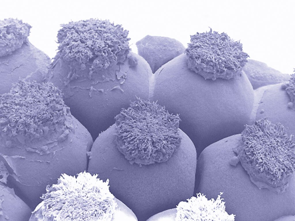
Nearly mature Aroid female flowers. The “fluffy” pad on each flower is the stigmatic surface that will receive pollen.
In Hibiscus rosa-sinensis, double flowers produce a number of stamens as well as petaloid floral organs on a donut-shaped meristematic ring normally designated exclusively for stamen production in typical flowers. In both systems, I want to determine if there are general relationships that characterize the substitution of one type of organ by another to uncover potential developmental constraints and underlying mechanisms.