

Rebecca Michaels-Walker

Rebecca Michaels-Walker is the Content Marketing Specialist at Canadian Science Publishing.

What are your cells “saying”? Dr. Dylan Burger on the language of extracellular vesicles

September 8, 2026 | 10 minute read

Science fiction loves a good alien communication problem. In the 2016 film Arrival, humanity struggles to decipher an alien language composed of complex circular symbols. In the 2026 film Project Hail Mary, the astronaut Ryland Grace initially misinterprets the capsules sent by the alien Eridians, only later realizing that they’re a form of communication. These stories challenge us to imagine what communication could look like in other species and on other worlds. What if living systems communicate in ways we haven’t learned to recognize? What can a “message” look like?

Researchers are asking the same questions when studying the cells in our own bodies. Cells have long been known to transmit extracellular vesicles (EVs): membrane-bound packages filled with molecular cargo. For years, scientists thought that these vesicles were little more than cellular garbage bags, carrying waste material out of the cell to be flushed away. But, like Grace on the Hail Mary, scientists are rethinking the purpose of these packages and realizing that EVs are actually more like envelopes carrying messages between cells.
Researchers now know that the molecular “messages” EVs carry can change the behaviour of other cells, coordinating processes including immune responses, tissue repair and, notably, cellular aging. As science fiction writers dream of turning back time, researchers wonder whether EVs could effectively pause or reverse our cellular clocks, slowing aging or regenerating tissues.

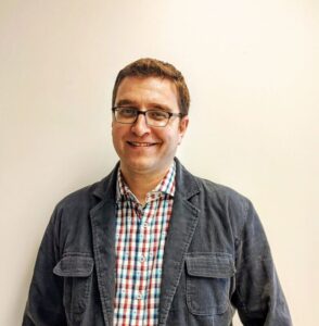
Dr. Dylan Burger, Co-Editor-in-Chief of the Canadian Journal of Physiology and Pharmacology, has built his career exploring these possibilities. Based at the University of Ottawa and the Ottawa Hospital Research Institute, his work has helped reveal how EVs contribute to aging, diabetes, and kidney disease. His team is pioneering the use of urinary EVs as early markers of kidney injury and is exploring how emerging technologies, like AI imaging tools, can help us understand new biological processes. In this interview, we talked to Dr. Burger about what cells are “saying” with their EVs, how these messages can promote health or cause disease, the possibility of slowing the aging process, and why science fiction continues to inspire the questions he asks as a researcher.
I think many of us imagine cells as self-contained units with their own jobs, busily working away with the help of a few close neighbours. You’ve spent much of your career studying EVs. What are EVs, and do they challenge this view?

EVs are pieces of the cell membrane that cells intentionally release into the extracellular environment. For instance, an endothelial cell in a blood vessel releases them into the blood, while an epithelial cell in the kidney might release them into the urinary space. Essentially, a portion of the cell’s outer shell is released, wrapped around some kind of contents. 

For a long time, people thought that cells were just releasing waste; vesicles were considered inert cellular debris. However, from the late 1990s to the mid-2000s, our understanding shifted: these vesicles are biologically active. This shift happened right around the time I was finishing my PhD. My background is in pharmacology and toxicology, particularly cell signaling and receptor biology. The idea that a piece of a cell could come off and trigger responses in target cells challenged what I learned in conventional pharmacology, which primarily focused on drug-receptor interactions for signaling pathway activation. 

This discovery fascinated me, prompting me to investigate why and how cells release these vesicles and if we could take advantage of them. The simplest example is that cells release a kind of larger EVs, called microparticles or microvesicles, when they’re stressed. These vesicles are highly pro-coagulant. If a blood vessel is damaged or infected, coagulation is essential as part of the bleeding response. Some studies have shown that these vesicles are up to 1,000 times more pro-coagulant than platelets. If a blood vessel is damaged or infected, coagulation is essential as part of the bleeding response. They also activate the immune system.  

Other vesicle classes, sometimes termed exosomes, transfer content from one cell to another, like envelopes carrying messages. If a cell wants to influence the behaviour of another cell—to cause it to grow or proliferate—it can release these vesicles, which transfer their content to the target cell.

Explore the latest advances in cardiovascular research from the 10th Annual Meeting of the European Section of the International Academy of Cardiovascular Sciences. READ THE COLLECTION

Do all cells release EVs?

As best we know, every cell that’s ever been studied releases EVs. That includes plant cells, bacteria cells, and human cells. The challenge with saying every cell does it is that, in theory, you have to prove that every cell does it! 

For example, in the body, it’s a lot easier to show that an endothelial cell lining a blood vessel or an immune cell in the blood releases EVs because you can take a blood sample and show that those vesicles came from those cells. Experimentally, it’s more difficult to show that a brain cell releases vesicles within the brain—it’s way harder to get into the brain to do testing than it is to take a blood sample.  

Additionally, some cells are better at releasing EVs than others. To my eternal suffering, endothelial cells don’t release very many EVs at all, even though I like studying them. In contrast, cells like macrophages or cancer cells tend to spit out a lot of EVs. We don’t always know why one cell is more active than others. However, as far as we’ve seen, pretty much every cell that’s ever been studied releases EVs.

Your work has shown that as we age, our cells change the “dialect” of the vesicles they release, shifting from smaller exosomes to larger ectosomes. Could you tell us a bit more about this discovery and what it explains about the process of aging? 

This line of inquiry has been a significant part of my lab’s work: what happens to EVs under certain conditions? We’ve also looked at diabetes, where we’ve shown that high glucose changes the type of EVs released and what those EVs do. Healthy endothelial cells release EVs that are relatively inert. However, under high glucose conditions, these EVs become far more pro-inflammatory and pro-coagulant.  

We’ve also investigated this in the context of aging. We were among the first to show that aging cells release vesicles that can induce aging in other cells. This creates a spiraling effect where healthy cells age because of signals from neighboring aging or “senescent” cells.  

We’ve shown that aging changes the types of vesicles released, favouring those formed off the blebs of the membrane (historically called ectosomes or microparticles). Conversely, there are fewer smaller vesicles originating from multivesicular bodies, known as exosomes. Exosomes from healthy cells generally appear beneficial, promoting cell growth and proliferation. However, when cells begin aging, they release vesicles that seem to damage neighboring cells. 

Canadian Science Publishing is a proud partner of the Society of Toxicology of Canada (STC).

Could we eventually learn to “re-tune” these messages to improve our health? 

One of the goals of my research program is to see if we can shift this balance. If molecular signals drive cells toward producing or “blebbing” harmful vesicles rather than beneficial ones, can we alter those signals? If we can, does it change what we observe in aging? 

The observation that EVs kick off an aging spiral aligns with what we casually observe with aging, right? We see a gradual progression in people, and then it suddenly seems to progress faster and faster, doesn’t it? If we can slow that initial progression or prevent it from happening, then we can potentially delay the aging process.  

We’re currently investigating a class of drugs called senotherapeutics, which target senescent cells, to see whether they can alter vesicle release and behaviour.  

Another, more speculative area, which we haven’t yet explored, is the diverse ways people age. This is something we also observe casually in real life. Individuals can be classified as slow, healthy, ultra-healthy, or rapidly aging people. When we think about rapid agers, it’s often in the context of cardiovascular disease or cognitive decline. Do these people have inherent differences in how their cells form EVs? Exploring this, alongside our current research, might help us understand why people age differently and identify ways to prevent this process. While we might not be able to stop those cells that initially become senescent, we might be able to stop them from making their buddies senescent. Hopefully we can slow things down that way. This could be a really big deal, especially for people suffering from cognitive decline or other progressive conditions. 

You’ve also expressed interest in how EVs could be used to regenerate tissues, which is kind of like the opposite of aging—turning back time. Can you tell us more about that? 

It all comes back to the main idea that a cell releases different types of EVs depending on what’s going on around it. If things are going well, some cells release beneficial, anti-inflammatory EVs that protect neighbouring cells and inhibit cell death. These are a big area of interest in regenerative medicine, which is one of the key research focuses at the Ottawa Hospital. 

I’ve collaborated with a number of researchers who have shown that if you treat a tissue with stem cell therapy, the stem cells disappear very quickly but their benefits remain. Many of those benefits can be replicated by treating tissues with vesicles collected from the stem cell cultures, suggesting that a lot of the stem cells’ power comes from their EVs. This is really appealing because it means maybe we could eliminate cell injection, which could reduce the risk of graft-versus-host disease. This is a significant global research area, with clinical trials progressing to use cell-derived vesicles for protection. 

My focus, however, is on preventing the body from producing harmful EVs and promoting the production of good ones. This aligns with my roots in pharmacology: instead of administering EVs, can we “trick” the body into producing healthier ones?

 The Canadian Journal of Physiology and Pharmacology has a new and expanded scope.

One of your lab’s big breakthroughs was identifying specific EVs (podocytes) in urine as markers of kidney injury. How might this discovery change the diagnosis and treatment of diabetic kidney disease? 

A cell’s EVs are like snapshots of its health. Examining them in the urine allows us to determine the current state of kidney cells. We initially looked at a large population of microparticles, which form under stress, and found them elevated in the kidney when there’s damage to its filtration system. 

Kidneys are your body’s vital filtration system. They filter your blood and remove waste through the urine. When the kidneys are damaged and can’t filter effectively, we call it kidney disease. The major cause of kidney disease is diabetes, and the first sign of diabetic kidney disease is generally protein leaking into the urine; the kidneys have been damaged and can no longer filter all the protein out. Over time, kidney function continues to decline and symptoms start to become evident.  

What excited us is that we see vesicles in the urine before we ever see protein. When protein begins to appear in the urine, irreversible damage has already occurred. The podocytes, the kidney’s filtering cells, have been damaged and they don’t recover sufficiently even with rapid treatment. If we can detect damage earlier, before it’s irreversible, we might be able to intervene and protect those cells. This is why urinary are particularly exciting for us. 

We’re currently working on developing a screening test that translates well to the clinic, but it’s been challenging. Most tests for studying EVs were designed for understanding biology in a research lab, not for rapid industrial use across clinical labs. 

People often underestimate the impact of kidney failure. I think most people understand that if you have cancer, you’re going to be in the hospital getting chemotherapy on a regular basis. I don’t think they realize that if your kidneys fail, you’re going to be in the hospital three times a week for dialysis for the rest of your life, unless you can get a kidney transplant. When you consider that 1 in 10 Canadians are impacted by kidney disease, that’s a huge burden for thousands of individuals and an enormous cost to the healthcare system. 

That’s why funding this type of research is so important. With more groups comparing different approaches to studying EVs in urine, and we’d find a method suited for clinical testing much faster. I’d love to see a pipeline where multiple labs simultaneously explore different technologies, so we can rapidly identify the most effective technologies and accelerate their translation into the clinic. 

A newer frontier for your lab is the study of Neutrophil Extracellular Traps (NETs). What are NETs, and how are you using AI to study them? 

Neutrophil extracellular traps are a relatively newly discovered and described mechanism our immune system uses to fight bacteria. Neutrophils (a kind of immune cell) spit out webs of their own DNA mixed with some enzymes that damage bacteria, such as myeloperoxidase. 

It really is a trap, and it really is a net. It’s one of the best scientific names that I’ve ever heard, because it’s exactly what it sounds like. The web of DNA traps the bacteria, preventing it from moving away, and then the neutrophil kills it with those bactericidal proteins. 

While NETs are fascinating in the context of immunology, I’m not an immunologist—I’m a vascular biologist by training. I became interested in what happens when NETs are dysregulated. This can occur in chronic diseases like kidney disease and diabetes. 

The challenge is that the tools for studying NETs aren’t always very good. My PhD student, Chloe, discovered this during her research. We developed our own tools, including an AI-powered imaging protocol to rapidly screen for NET formation. This is incredibly useful because the human eye isn’t very good at doing this, especially not quickly and in large volumes. This makes AI-guided analysis ideal. We’re now leveraging this technology to connect it to some of our favourite questions, like: How does aging affect NET formation? Do EVs influence NET formation?

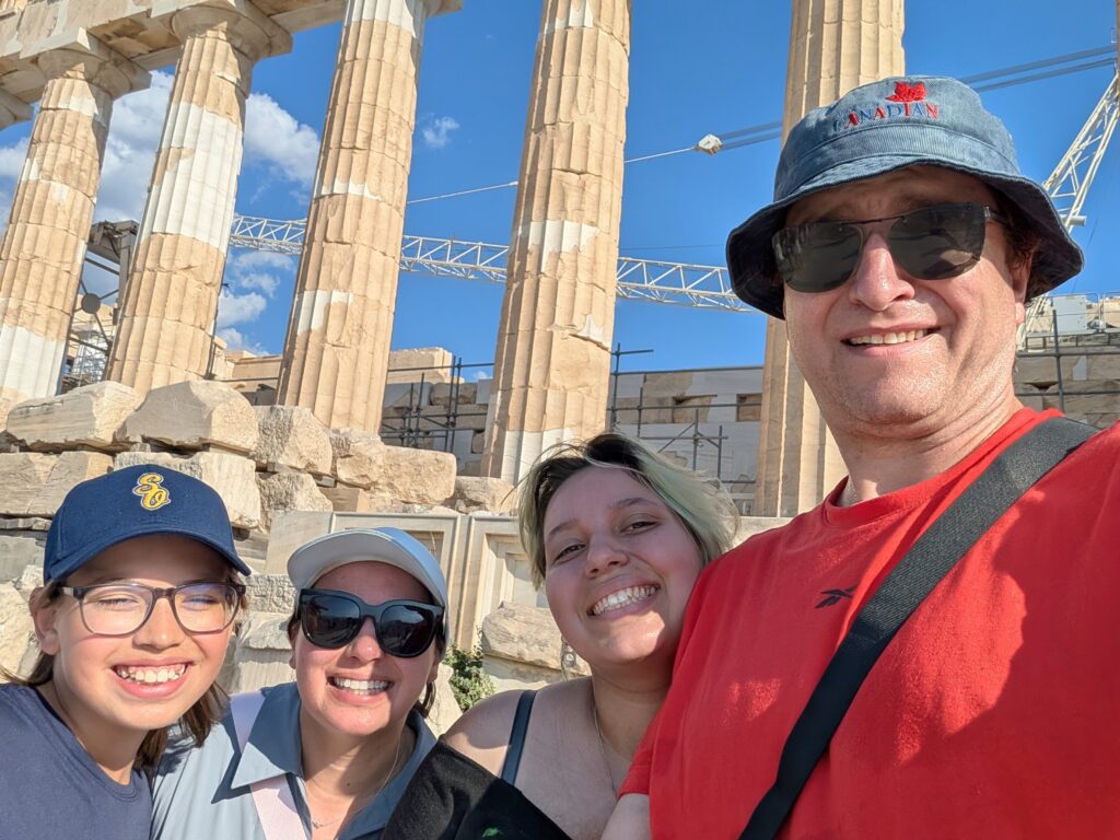
Dr. Burger and his family.

With “Project Hail Mary” and the NASA mission this year, a lot of people have renewed excitement about how sci-fi can inspire “real” science. Are you a fan of the genre? 

You know I am! I think one of the things I love about the sci-fi genre, particularly Andy Weir’s work—Project Hail Mary, The Martian, Artemis—but also Michael Crichton’s work, is that it’s a really good story so you don’t immediately tune it out, and it’s rooted in real science, so you don’t immediately dismiss it as far-fetched. The kind of sci-fi that I really like is rooted in real science and looks at it from unusual directions. Project Hail Mary explores whether life can exist that is not carbon-based, for example. I think sometimes in science, it’s easy to focus on the negative—what doesn’t work, what doesn’t make sense, what isn’t possible. But I think sometimes we need to be regularly interacting and thinking in ways that ask, “Okay, given this, is this possible? What could happen?”  

Of course, science fiction writers don’t always get it right. But at the same time, it’s challenging you to think about how things work in unique ways and makes you consider new possibilities. That’s why it’s my genre. I’ve gotten my kids into it too. Their favourite book is now Project Hail Mary. They were more excited about the movie than I was! 

Your academic journey began with an interest in psychology, yet you’ve said that within five minutes of stepping into a fourth-year undergraduate lab, you knew discovery research was your calling. What did you experience in that lab that fundamentally altered your life’s trajectory? 

Well, I will say it wasn’t as much a transition as a prioritization. My undergrad was a lesson in how to avoid making a decision! I wasn’t sure if I wanted to study biology or sociology, so I started in biology because it was easier to switch from biology to sociology than the other way around. Then I moved into pharmacology because it had an extra elective compared to other biology disciplines, which allowed me to keep minoring in psychology. Then I joined a research lab, and I knew very quickly that’s what I wanted to do. A number of things persuaded me: the opportunity to be around smart people, discussing, speculating, and going back and forth between ideas. It also fulfilled my desire to explore the unknown, which has been with me since childhood. It was also the collegiality—a lab is a community. Even when your own work is incremental, it feels like you’re part of something bigger. Together, you’re building science. 

Rebecca Michaels-Walker

Rebecca Michaels-Walker is the Content Marketing Specialist at Canadian Science Publishing.